Case 4: SNAP™ Therapy Diabetic Foot Wound
Patient information:
68-year old male
68-year old male
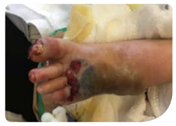
Wound history:
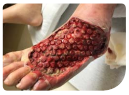
Trauma to dorsal foot from door.
Trauma to dorsal foot from door.
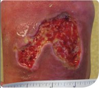
Dimensons:
Prior to SNAP™ Therapy, the wound measured 70mm x 54mm with a depth of 4mm without undermining.
Prior to SNAP™ Therapy, the wound measured 70mm x 54mm with a depth of 4mm without undermining.
Comorbidities:
Diabetes Mellitus, Smoker, Peripheral Vascular Disease, Coronary Artery Disease, COPD, Hypertension, Hyperlipidemia.
Diabetes Mellitus, Smoker, Peripheral Vascular Disease, Coronary Artery Disease, COPD, Hypertension, Hyperlipidemia.
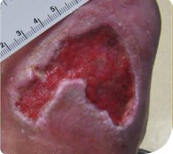
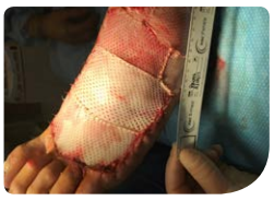
SNAP™ therapy treatment:
The patient was treated with SNAP™ Therapy for 3 weeks until full granulation of the wound bed was achieved. Then SNAP™ Therapy was used in conjunction with a cellular tissue product for an additional 5 weeks.
The patient was treated with SNAP™ Therapy for 3 weeks until full granulation of the wound bed was achieved. Then SNAP™ Therapy was used in conjunction with a cellular tissue product for an additional 5 weeks.
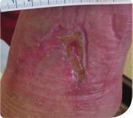
Outcome:
Wound closure was achieved at 9 weeks post-initiation of SNAP™ Therapy.
Wound closure was achieved at 9 weeks post-initiation of SNAP™ Therapy.